
Advances in 3D bioprinting technology could pave the way for new approaches to heart disease treatment and disease modeling.
Researchers at the University of Galway have announced a significant advance in the field of 3D bioprinting – successfully fabricating functional human heart tissue. Their research, which has been published in Advanced Functional Materials, describes the development of bioprinted hydrogels that mimic the mechanical, electrical and biochemical environment of the heart – an essential stage in creating viable tissue for regenerative applications and drug development and one that can be heralded as a step forward in the development of patient-specific cardiac therapies.
Longevity.Technology: Heart disease remains one of the leading causes of mortality worldwide; with a significant shortage of donor hearts, the demand for alternative solutions has never been more pressing. The creation of functional heart tissue offers the potential to address this unmet need, offering both a way to advance research into cardiac conditions and a future source of therapeutic options.
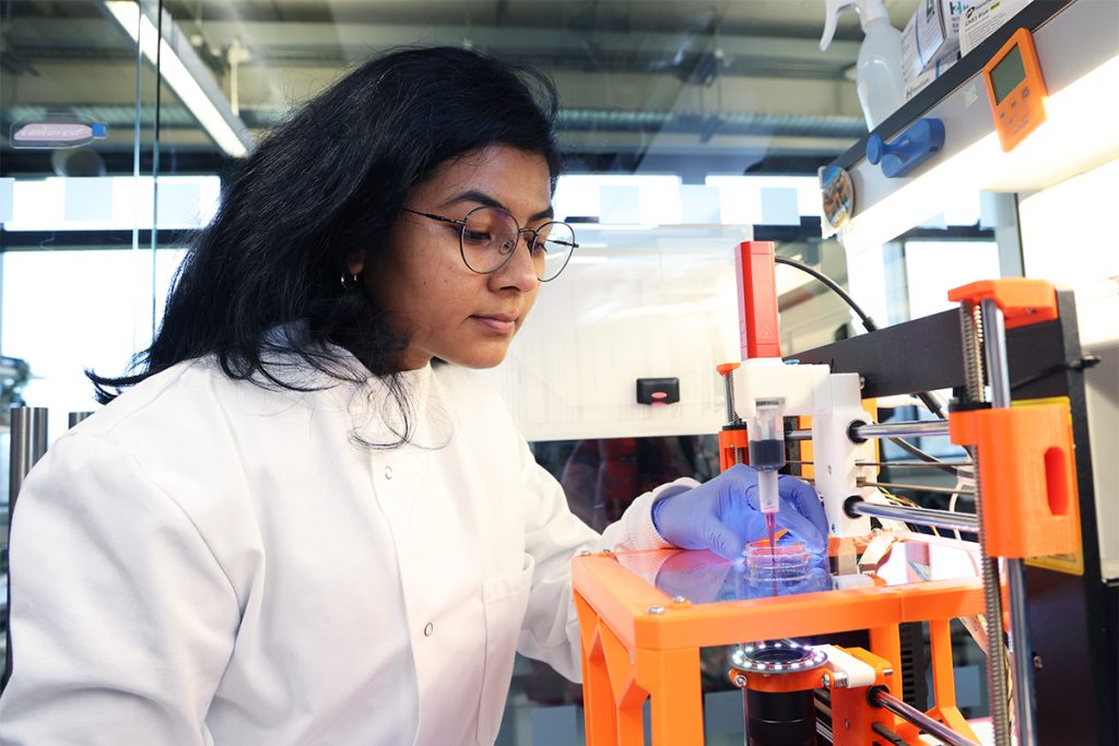
The team’s approach centered on the use of extrusion-based bioprinting techniques to create structured hydrogels designed to support cardiac cell growth [1]. The bioink used closely mimicked the properties of the extracellular matrix, enabling the creation of tissue constructs that demonstrated both mechanical integrity and biological function.
The bioprinted tissue demonstrated synchronized contractions as well as compatibility with long-term cellular survival – these findings suggest that bioprinting could eventually lead to patient-specific therapies for cardiovascular diseases.
A focus on functional outcomes
The breakthrough lies not only in the ability to replicate heart tissue structures but also in ensuring their functionality.
Conventional bioprinting approaches often focus on replicating the final form of organs, such as the heart, without accounting for the dynamic transformations that occur during embryonic development. For example, while the heart begins as a straightforward tube, over time it bends and twists into a complex four-chambered structure – these dynamic shape-morphing changes play a critical role in the growth and specialization of heart cells.
To improve on convention, the Galway researchers introduced a innovative bioprinting method that incorporates these essential shape-changing behaviours.
Ankita Pramanick, lead author of the study and CÚRAM PhD Candidate at University of Galway, said: “Our work introduces a novel platform, using embedded bioprinting to bioprint tissues that undergo programmable and predictable 4D shape-morphing driven by cell-generated forces. Using this new process, we found that shape-morphing improved the structural and functional maturity of bioprinted heart tissues [2].”
The bioprinted constructs were evaluated for their contractile behavior, cell viability and molecular expression; results demonstrated that the tissue constructs could contract synchronously, a hallmark of functional cardiac tissue, and this ability is critical for applications in regenerative medicine and for creating accurate models to study diseases such as cardiomyopathies.
The research demonstrated that cell-generated forces could drive the morphing of bioprinted tissues, with the extent of these shape transformations being influenced by factors like the initial print geometry and bioink stiffness [1]. Morphing was shown to play a key role in shaping cell alignment and improving the tissues’ contractile properties. Additionally, the research team created a computational model capable of predicting tissue morphing behavior.
Professor Andrew Daly, Associate Professor in Biomedical Engineering and CÚRAM funded investigator and principal investigator on the project, said: “Our research shows that by allowing bioprinted heart tissues to undergo shape-morphing, they start to beat stronger and faster. The limited maturity of bioprinted tissues has been a major challenge in the field, so this was an exciting result for us. This allows us to create more advanced bioprinted heart tissue, with the ability to mature in a laboratory setting, better replicating adult human heart structure. We are excited to build on this shape-morphing approach in our ongoing European Research Council project, which is focused on developmentally-inspired bioprinting [2].”
Expanding possibilities for cardiac research and therapy
One of the immediate applications of the bioprinted heart tissue is its potential use in drug screening. Current models for testing cardiac drugs often rely on animal tissues, which do not fully replicate human cardiac biology; the ability to produce human tissue constructs offers a more accurate and ethical alternative, allowing pharmaceutical companies to test the safety and efficacy of treatments with greater precision.
In the longer term, the technology could contribute to solving the organ shortage crisis. While full organ bioprinting still remains a distant goal, advancements in the fabrication of functional tissue like these are a vital precursor. The researchers emphasize that scalability and reproducibility will be key challenges as they move forward – particularly in adapting the technology for clinical applications.
The challenges of clinical translation
Despite the promising outcomes, there remain significant hurdles to overcome before bioprinted heart tissue can be used in a therapeutic setting. Ensuring the integration of bioprinted constructs with native tissues, scaling up production to meet clinical demands and addressing regulatory hurdles will all require further research and development.
“We are still a long way away from bioprinting functional tissue that could be implanted in humans, and future work will need to explore how we can scale our bioprinting approach to human-scale hearts,” said Daly.
“We will need to integrate blood vessels to keep such large constructs alive in the lab, but ultimately, this breakthrough brings us closer to generating functional bioprinted organs, which would have broad applications in cardiovascular medicine [2].”
Implications beyond cardiology
As well as developing a novel bioprinting platform, the team was able to simulate shape-changing behaviors at both the cell and tissue levels using models that mimic how fibers within the tissue rearrange themselves [1]. This ability to design, predict, and program 4D shape-morphing in bioprinted tissues has the potential to transform organ engineering. Instead of focusing solely on recreating the final shape of an organ, this approach emphasizes mimicking the natural developmental processes that guide its form, structure and function. This shift opens exciting new possibilities in organ bioprinting.
Although this study focuses on heart tissue, the techniques developed have broader implications for the field of regenerative medicine. Similar approaches could be applied to create functional tissues for other organs, opening the door to advances in the treatment of diseases ranging from liver failure to diabetes. The interdisciplinary nature of this work, combining cutting-edge materials and biological science, highlights the potential of 3D bioprinting as a transformative technology in medicine.
[1] https://advanced.onlinelibrary.wiley.com/doi/10.1002/adfm.202414559#adfm202414559-bib-0004
[2] https://tinyurl.com/4b836r2u

